Services
- Cellular and Tissue Morphology
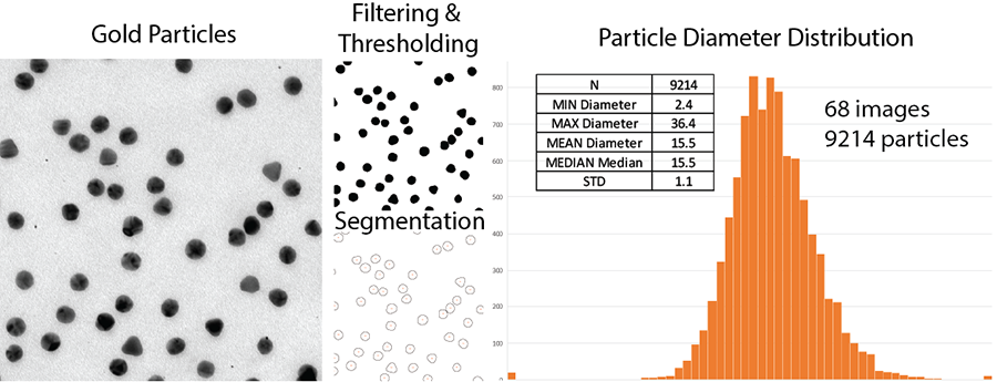
- Nanoparticle Imaging & Analysis
- Immunolabelling for Protein Localization
- CLEM: Providing Structural Context to Fluorescence
- Electron Tomography
Viruses, proteins, vesicles, suspensions, or nanoparticles are placed on the EM grids and can be observed directly by electron microscopy. For better results, samples are negatively stained.
This is the most direct visualization of a purified sample. It informs on preparation concentration and homogeneity and enables structural characterization of the particles.
This method can be combined with immunolabeling to verify the presence and localization of a protein of interest.
We can perform the staining and imaging of your samples or train you to do it.
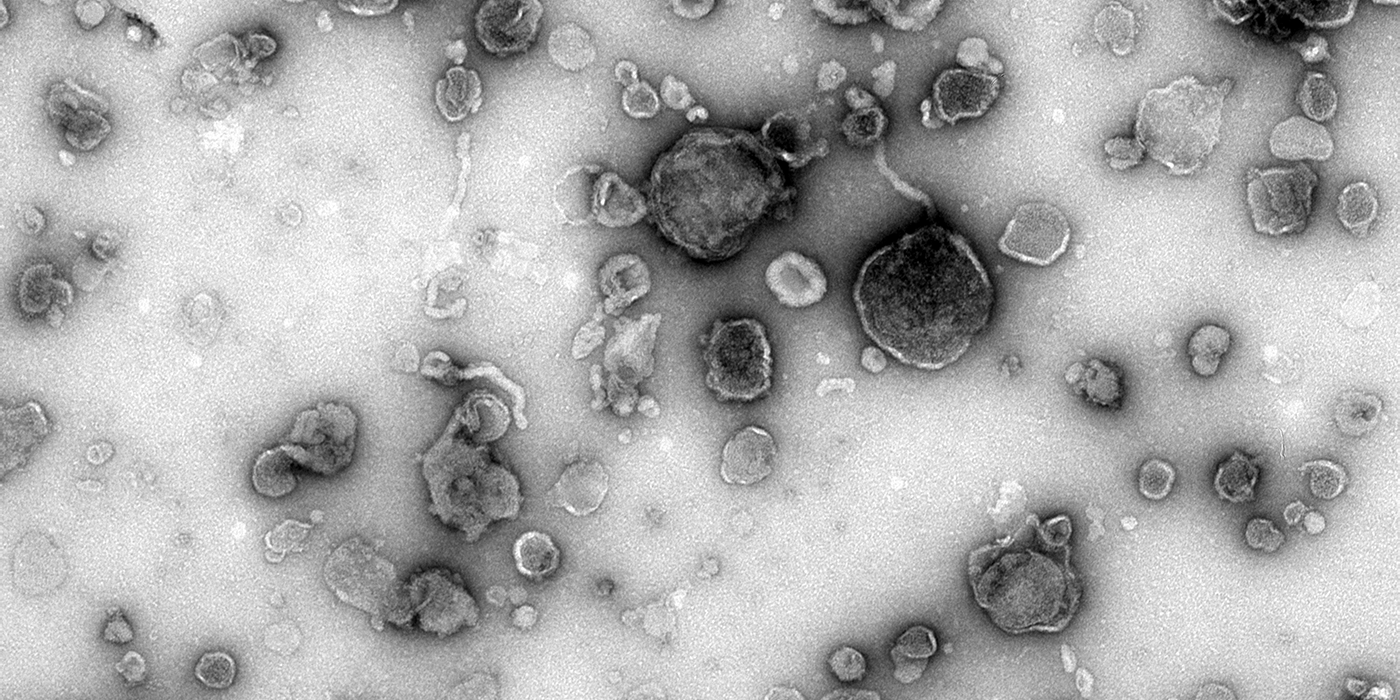
Extracellular Vesicles-EV
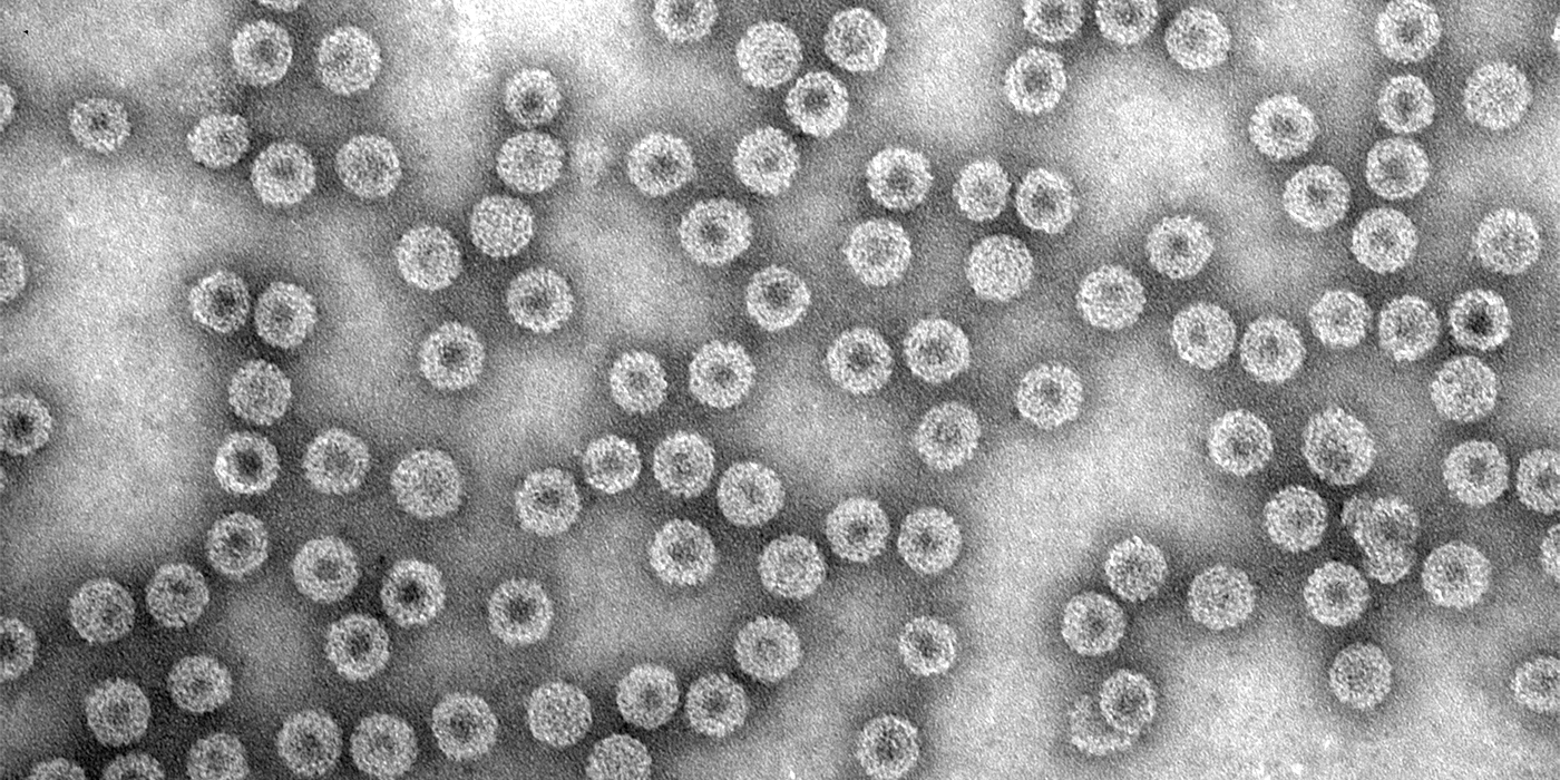
Virus
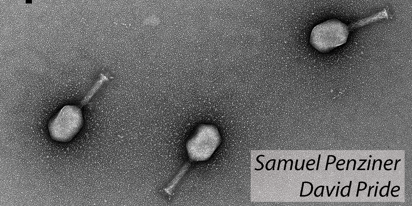
Bacteriophages
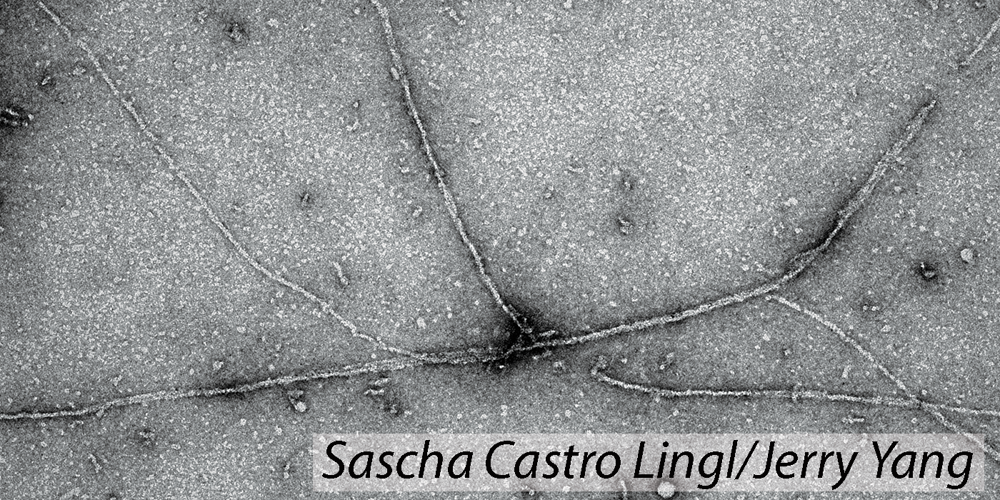
In vitro Polymerized Actin
We can assist you with the measurements of the nanoparticle features, such as: